Do Orthopedic Surgeons Have Their Own Ray Machines?
Orthopedic surgeons use a large number of specialized tools to perform their job. Besides the tools that are used in surgery, there are also a lot of tools that are used by orthopedic surgeons, but that “belong” to other specialties (e.g., radiologists). One of these is the X-ray machine.
- Ray Machines
- Orthopedic surgeons don’t have their own x-ray machines
- What machines do orthopedic surgeons use?
- .box-4-multi-105{border:none !important;display:block !important;float:none;line-height:0px;margin-bottom:15px !important;margin-left:0px !important;margin-right:0px !important;margin-top:15px !important;max-width:100% !important;min-height:250px;min-width:250px;padding:0;text-align:center !important;}Stryker Navigation Systems
- The Zimmer OrthoPilot
- Smith & Nephew NAVIO Surgical System
- The DePuy Synthes MAKO System
- Blue Belt Technologies Navio System
- Are orthopedic surgeons exposed to radiation?
- Wearing protective gear
- What is a ray in orthopedics?
- Who is qualified to read an x-ray?
- Medical imaging scientists
- Medical physicists
- Diagnostic radiographers
- Sonographers
- Nuclear medicine practitioners
- Can orthopedic surgeon read MRI?
- Do radiologists perform surgery?
- Are radiologists real doctors?
- What is the difference between radiology and radiography?
- How many years does it take to become a radiologist?
- Do radiologists read MRI?
- What are the five most common errors in radiology?
- Lack of communication of helpful information to the receiving clinician:
- Improper discussion or collaboration among radiologists:
- Overly focused pattern recognition reading a case in isolation (what do I see):
- Failure to communicate a diagnosis, incomplete or delayed diagnosis, or wrong diagnosis communicated to the patient.
- What do radiologists do all day?
- Answer questions
- Carefully study images
- Measure, measure, and re-measure
- Collaborate with other caregivers
- Talk to patients
- Educate the next generation of radiologists
- Do radiologists see patients?
- How often do radiologists make mistakes?
- Can a neurosurgeon read an MRI?
- Can you sue a radiologist for misdiagnosis?
- Medical malpractice
- Are chiropractors trained to read MRI?
- How often is an MRI wrong?
- Do radiologists make diagnosis?
- What happens if a radiologist makes a mistake?
- Get a second opinion
- Decide whether to pursue legal action
- Start the process of filing a lawsuit
- You can fight back if you think a radiologist made a mistake
- Conclusion
Ray Machines
Doctors who specialize in orthopedics use the same x-ray machines as other medical professionals, and they don’t have their own. Orthopedic surgeons are general surgeons who specialize in the treatment of bones, joints, and ligaments. They typically work closely with radiologists or radiographers to order diagnostic tests like x-rays when necessary.
The fact that orthopedic surgeons don’t have their own ray machines is a good thing because it means that your doctor won’t need to spend extra time waiting for an appointment to get an x-ray done if you need one.
Orthopedic surgeons don’t have their own x-ray machines
Orthopedic surgeons don’t have their own x-ray machines. They use x-ray machines in hospitals and are trained to use them. Many orthopedic surgeons also work as radiologists, but they still don’t have their own x-rays.
Orthopedic surgeons will use medical imaging like X-rays to diagnose injuries and diseases of the bones or muscles.
The most common conditions that require an orthopedic surgeon are arthritis or joint pain associated with age or overuse of a joint such as tennis elbow or carpal tunnel syndrome.
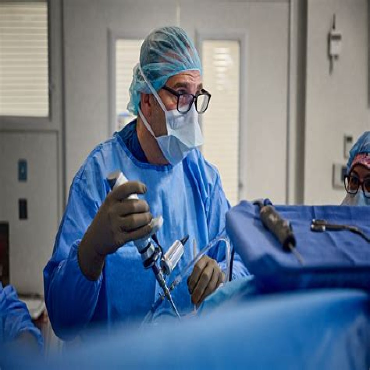
What machines do orthopedic surgeons use?
Stryker Navigation Systems
Stryker Navigation Systems are used to assist with knee surgery. They help surgeons track the knee joint’s position and make accurate cuts and measurements.
The Zimmer OrthoPilot
The Zimmer OrthoPilot is a robotic system used by orthopedic surgeons to guide knee surgery. It looks like a small cart with two robotic arms; one arm is attached to the patient’s leg, while the other guides the surgeon as they perform surgery. This device improves accuracy during knee surgeries and reduces risks for patients and doctors.
Smith & Nephew NAVIO Surgical System
The Smith & Nephew NAVIO Surgical System is a device used for hip and knee replacement surgery. It’s used to plan the surgery, guide surgeons, visualize what they’re doing in 3D, and control robotic arms that perform specific movements during the operation.
The DePuy Synthes MAKO System
The MAKO System is a surgical navigation system that uses a camera and a computer to help surgeons perform surgeries. The MAKO system performs hip, knee, and shoulder surgeries.
Blue Belt Technologies Navio System
The Blue Belt Technologies Navio System is a surgical navigation system used in orthopedic surgery. The system guides the surgeon during hip and knee replacement surgery, which helps the surgeon find the correct position for the surgery. This can help reduce pain and postoperative complications for patients.
Are orthopedic surgeons exposed to radiation?
Orthopedic surgeons are one of the groups most exposed to radiation in the hospital.
This is because they perform several different types of surgery, which may have higher levels of ionizing radiation, such as computer tomography (CT) scans or fluoroscopy. In addition, orthopedic surgeons often work with radiation oncologists, who specialize in using high doses of radiation to treat cancerous tumors and cancers.
Orthopedic surgeons get a radiation exposure equivalent to 1,800 chest X-rays per year. On average, orthopedic surgeons get a radiation exposure equivalent to 1,800 chest X-rays per year. This number is calculated using the National Council on Radiation Protection and Measurements (NCRP) formula for radiation exposure.
Wearing protective gear
Of course, wearing protective gear will not protect you from all radiation exposure. However, it can help keep your exposure within reasonable levels. This will be especially important if you are working with high-energy radiation sources or radioactive materials. Proper use of tools and equipment and minimizing their service is also essential in protecting yourself from radiation.
A lead apron can protect the stomach, hips, pelvis, and thighs. The leaded apron should not be worn over clothing or other protective garments such as scrubs or lab coats.
A lead glove protects the hands from radiation when handling radioactive materials such as needles and syringes. The lead should cover the entire hand, including fingers and thumb, with no gaps between fingers.
What is a ray in orthopedics?
A ray is a set of bones in the hand, foot, and leg. There are five rays on each side of your body: metacarpals (5) in your hands, metatarsals (14) in your feet, and phalanges (27) in your toes.
The bones that ligaments and joints connect make up rays. These connections allow you to move with ease when walking or running.
If one of these connections becomes damaged or broken, it can cause pain when moving certain parts of your body. The most common types of injuries affecting rays include fractures and amputations.
The term “ray” was initially used to describe the rays of light that shone from a light source.
Rays are found in many body areas, including the hand, foot, leg, and skull. The spine also contains rays that allow it to bend when moving your body without breaking. Ray-related surgeries are often performed on the hand, foot, or leg bones.
When a surgeon performs a ray amputation surgery on their patient’s hand, they remove one or more of these rays so their muscles can no longer use them—this is because it helps prevent infection from spreading through the body.
A ray amputation surgery is typically done only when other treatments have failed to stop recurring infections in an area where there may be multiple fractures (such as when someone breaks their arm).
Who is qualified to read an x-ray?
A radiologist is a doctor who reads X-rays, ultrasounds, and other imaging exams. Radiologists are qualified to read these tests because they have a medical degree and rigorous training in interpreting medical images.
Medical imaging scientists
You may hear the term “medical imaging scientist” used when referring to someone who works with medical images. These individuals work in hospitals and clinics and often hold the title of a radiographer or radiologic technologist.
Medical physicists
Medical physicists are the scientists who use physics to help diagnose and treat diseases. They’re not medical doctors, but they can apply their knowledge of physics to help diagnose diseases.
Diagnostic radiographers
A diagnostic radiographer is a healthcare professional trained to interpret x-rays and other images to diagnose health problems. They also operate X-ray machines and other imaging equipment.
Sonographers
Sonographers are also known as diagnostic medical sonographers. They use diagnostic ultrasound to produce images of the human body and work with radiologists and other physicians.
Nuclear medicine practitioners
Nuclear medicine practitioners are medical doctors who undergo training in nuclear medicine. They use radioactive isotopes to diagnose and treat disease but are not required to be board-certified in anesthesiology, emergency medicine, or pathology.
Can orthopedic surgeon read MRI?
Orthopedic surgeons and radiologists are the two types of doctors who can read your MRI. These two doctors have different areas of expertise.
The radiologist is a doctor who specializes in reading medical imaging scans. At the same time, the orthopedic surgeon is a doctor who treats issues with the musculoskeletal system, which includes bones and joints.
Although radiologists are trained to read MRIs, only orthopedic surgeons can apply the results of your MRI scan to a plan for treatment for any problems with your bones or joints.
Do radiologists perform surgery?
No. Radiologists are medical professionals who diagnose and treat diseases using medical imaging, such as x-rays, CT scans, MRIs, and ultrasounds. They work with patients to determine the procedure needed for the best diagnosis and outcome.
Specifically, in radiology, you will help patients understand their disease process through imaging so they can make informed decisions about their care plan. The vast majority of radiologists do not perform surgery at all.
Are radiologists real doctors?
Yes, they are. You probably think that radiologists are just people who read X-rays and CT scans. But they’re doctors with a specialty in radiology, meaning they have the same training and responsibilities as other MDs.
Radiologists have completed four years of medical school and another three to four years of residency before obtaining their specialization in radiology.
They are also licensed to practice medicine, which means they can diagnose and treat patients with any disease or condition.
What is the difference between radiology and radiography?
Radiology is a branch of medicine that uses imaging techniques and equipment to diagnose, monitor, and treat various medical conditions.
Radiography is an imaging technique that uses X-rays to get information about internal body structures or tissues. Although radiology and radiography are related, there is a differences between them.
How many years does it take to become a radiologist?
On average, it takes 13 years to become a radiologist. Perhaps the earliest thing to consider is the long it will take you to complete your undergraduate education. After that, it’s time to get into medical school and begin your training in radiology.
At a minimum, you’ll need four years of undergraduate study before entering medical school. Isuppose you want to become a radiologist specializing in nuclear medicine or diagnostic imaging (CT/MRI). In that case, this can add another year or two to your time frame for becoming a doctor.
The medical school typically lasts four years as well; after completing that stage of your education and getting licensed by taking the USMLE exams (a requirement for all MDs). It’s time for internship—the first year where medical students are on their own with patients under supervision from attending physicians—followed by residency training lasting anywhere from 4-5 years depending on what specialty area you choose (anesthesiology, emergency medicine).
Do radiologists read MRI?
Yes, radiologists do read MRI. Radiologists are physicians that specialize in diagnosing and treating diseases using medical images.
Radiologists are the specialists to diagnose and treat diseases using medical imaging technologies like x-rays, Mammography, PET, MRI, CT scan, ultrasound, and Nuclear medicine imaging.
What are the five most common errors in radiology?
A misdiagnosis can have serious consequences, even if it’s only a matter of weeks or months. Here are some common causes of misdiagnosis in radiology:
The patient has a rare disease; the radiologist doesn’t know about it and doesn’t ask for help from another expert.
The radiologist has poor knowledge about how common diseases present in specific populations.
Lack of communication of helpful information to the receiving clinician:
Without this information, the treating clinician is forced to start essentially from scratch on their patient. This can lead to delays in treatment and an increased likelihood of errors.
Improper discussion or collaboration among radiologists:
If more than one radiologist is involved in reading an image, they must communicate effectively and reach a consensus about the findings.
Overly focused pattern recognition reading a case in isolation (what do I see):
What do we expect? We expect to see patterns, abnormalities, signs of disease, and the correct answer. It’s not always that simple, though.
Patients are individuals, and their differences can affect how they present on imaging. How is that relevant? Well, if you think about patterns and expected findings, you may miss something important because it doesn’t fit into your pre-defined box.
We don’t want to miss anything! So what can we do instead?
Failure to communicate a diagnosis, incomplete or delayed diagnosis, or wrong diagnosis communicated to the patient.
This is a big one. When communicating a diagnosis, it’s essential to be clear and concise, not just with the patient.
What do radiologists do all day?
Answer questions
Radiologists are doctors who specialize in interpreting X-rays, CT, and MRI scans. They work with patients and their doctors to provide the best possible care.
For example, a radiologist may read an X-ray of a patient’s spine to determine whether there is anything unusual about it or whether the patient needs additional imaging studies.
Carefully study images
A radiologist’s job is to study images and diagnose abnormalities in the human body carefully. They are trained to look for things that don’t seem normal, such as abnormal growth or a tumor.
Measure, measure, and re-measure
One of the most fundamental parts of radiologist work is measuring. Whether it’s measuring the size of a tumor, blood vessel, bone, brain, or heart, radiologists need to know precisely how large an organ is to ensure they’re getting an accurate picture of its health.
Collaborate with other caregivers
Radiologists are constantly collaborating with other healthcare professionals. They need to work with patients, their peers and families, and even the friends of their patients. It can be a lot for one person to handle.
Talk to patients
When you go for an MRI or CT scan, your image appears on a screen. A radiologist is the first doctor to see the results of a scan. They explain what they know to you and will often offer advice on what might need further tests, like a ultrasound or biopsy.
Educate the next generation of radiologists
It’s important to note that radiologists also play an essential role in educating the next generation of radiologists. They teach medical students, residents, and other medical professionals who might not have had much exposure to radiology before.
Do radiologists see patients?
There is a common misconception that radiologists don’t see patients. The truth is that radiologists sometimes see patients, but not in the same way as other physicians like your family doctor.
Radiologists are medical professionals who specialize in interpreting medical images and producing reports for other physicians to use when treating patients.
Radiologists don’t treat you directly or provide medical advice. Instead, they work very closely with other physicians (such as surgeons) to help them provide you with the best possible care by providing them with information about your condition from a different perspective than their doctors have based on direct observation of your symptoms and examination findings.
How often do radiologists make mistakes?
Radiologists and physicians make medical errors at a low rate of only 1%.
To be clear, radiologists are not infallible and are human and make mistakes, but the error rate is low. Fortunately the rate has been declining over time and is not significantly different from other medical fields.
The National Patient Safety Foundation reports that “medical errors” account for 98,000 deaths yearly. While there is no doubt that mistakes happen from time to time in radiology (and other specialties), it’s essential to keep things in perspective when considering the scope of such errors on an annual basis across all specialties within medicine.
Can a neurosurgeon read an MRI?
Neurosurgeons can read MRI images, though they often use radiologists to double-check complex cases. Neurosurgeons can read MRI images, though they often use radiologists to double-check complex cases.
A neurosurgeon can indeed read an MRI image if they have been trained to do so. However, there are many different types of doctors who read MRIs: radiologists, neuroradiologists (neurologists who specialize in reading MRIs), and neuropathologists (doctors who study tissue samples). Neurosurgeons usually look at the whole brain, while neuropathologists focus on looking at individual tissues like blood vessels and neurons.
Can you sue a radiologist for misdiagnosis?
Misdiagnosis in and of itself isn’t grounds to sue a doctor.
Misdiagnosis is not the same as negligence, which is the failure to do what a reasonably competent doctor would have done in the same situation.
It’s also not malpractice, which means professional misconduct that harms a patient. Misdiagnosis isn’t grounds for suing a doctor or even an individual radiologist—it’s just part of being human and why getting second opinions is essential.
Medical malpractice
If a radiologist misreads your medical imaging, your state’s laws will determine whether you can sue for medical malpractice. To file a lawsuit, you must prove that the doctor was negligent and that their negligence resulted in harm to you.
If a radiologist is negligent, then a patient may be able to recover damages for pain and suffering and economic losses related to the misdiagnosis.
Are chiropractors trained to read MRI?
Some chiropractors do interpret MRIs. They receive specialized training as part of their degree program and may even use the scans to help diagnose and treat patients.
It’s important to know what kind of training your chiropractor has had.
Many chiropractors are not trained to read MRIs, so don’t assume that all chiropractors can read MRIs.
You may also want to ask about their training related to the spine and other types of injuries. The kind of work they do depends on how much education and experience they have in the field – there are many different types of doctors who treat back pain.
How often is an MRI wrong?
It’s important to remember that an MRI may not be completely accurate. About 10% of the time, an MRI result is wrong.
If your doctor tells you they’ve found something on your MRI, they might be wrong. It’s possible that there’s nothing wrong at all and everything is fine. Or maybe there IS something wrong, but it’s not what the radiologist thinks it is.
So try not to worry too much about what could happen if your scan shows something unusual; rather than worrying about unfortunate outcomes such as cancer or a brain tumor, keep in mind that there are many things an MRI can’t detect and even more reasons why it may not be accurate in some cases.
Do radiologists make diagnosis?
Yes. While it sounds like a simple question, the answer is not always straightforward. Radiologists are medical doctors who specialize in interpreting images produced by diagnostic imaging technology.
They are an essential part of the diagnostic process, but they are only one part.
The other parts include the patient, their family, and their referring physician. The radiologist can only interpret these images; they cannot diagnose your problem without more information from these sources.
They also cannot order any treatments for you or provide any care after a diagnosis has been made except for follow-up imaging studies if needed or recommended (for example, CT scans following surgery).
What happens if a radiologist makes a mistake?
Find out if a mistake was made
On the off chance that you think your primary care physician committed an error, it’s essential to request a duplicate of the report, to request a duplicate of the report, and this should include all relevant information about the test and its results. You will also want to get the name of the radiologist who read your X-rays and any other doctors involved in your case. If possible, note down their names and contact numbers so that you can ask for clarification if needed.
Get a second opinion
Getting a second opinion is one of the most common ways to ensure you’ve received the best possible care. If a radiologist’s interpretation of your scans differs from what you expected it to be, or if you are not satisfied with the results, you need to get an opinion from another radiologist.
Decide whether to pursue legal action
When you discover that your doctor has made a mistake, you may be tempted to pursue legal action against them. However, weighing the pros and cons is essential before deciding what course of action is suitable for you.
Start the process of filing a lawsuit
If you’ve been injured by a radiologist’s mistake, start the process of filing a lawsuit. You’ll want to file in the state where the radiologist works and lives. In some cases, you can also use it against multiple parties: the hospital that employed them, their insurance company, and even their medical provider or medical facility for negligent hiring practices.
You can fight back if you think a radiologist made a mistake
If you believe a radiologist made a mistake, the first step is to see if you can get them to admit it. Also, if the radiologist in question refuses to accept fault and provide compensation, your second step will be deciding whether or not to pursue legal action.
If you choose not to file a lawsuit and instead pursue other routes for resolving your medical malpractice claim (such as mediation), then your final step would be filing paperwork with the state’s Board of Medical Examiners and Health Department to start the process of filing a lawsuit.
Conclusion
There are many systems that orthopedic surgeons can use to perform surgery. Orthopedic surgeons will generally not be exposed to radiation during surgeries. This is because they do not perform X-rays. Ray-related surgeries are often performed on the hand, foot, or leg bones. An orthopedic or podiatric surgeon usually does Ray’s amputation surgery.
Hopefully, this article has given you a clearer understanding of the different job roles in radiography, what happens when a radiologist makes a mistake, common errors in radiology and how they fit into the team of healthcare professionals. It’s important to remember that all these roles are equally important.
